|
Green fluorescent protein TagGFP2
- Bright green fluorescence
- Monomeric protein with successful performance in fusions
- Fast maturation, high pH-stability and photostability
- Proven suitability to generate stably transfected cell lines
- Recommended for protein labeling and FRET applications
 |
TagGFP2 (scientific name mTagGFP) is the improved variant of TagGFP, the mutant of the Aequorea macrodactyla GFP-like protein [Xia et al., 2002, Subach et al., 2008]. TagGFP2 possesses bright green fluorescence with excitation/emission maxima at 483 and 506 nm, respectively.
TagGFP2 matures 1.6-fold faster than TagGFP and is characterized by the improved performance in fusions. Compared to EGFP, TagGFP2 provides about the same brightness of fluorescence but is significantly more pH stable. TagGFP2 is specially optimized for expression at 37°C.
Because of monomeric nature, TagGFP2 is mainly intended for protein localization studies and expression in long-term cell cultures. In FRET applications, TagGFP2 can be used as a donor for red fluorescent protein TagRFP or as an acceptor for blue fluorescent protein TagBFP.
|
Main properties
TagGFP2 normalized excitation (thin line) and emission (thick line) spectra.
Spectra viewer tool
Download TagGFP2 spectra (xls)
| | CHARACTERISTIC | |
|---|
* Brightness is a product of extinction coefficient and quantum yield, divided by 1000.
| | Molecular weight, kDa | 27 | | Polypeptide length, aa | 238 | | Fluorescence color | green | | Excitation maximum, nm | 483 | | Emission maximum, nm | 506 | | Quantum yield | 0.6 | | Extinction coefficient, M-1cm-1 | 56 500 | | Brightness* | 33.9 | | Brightness, % of EGFP | 105 | | Fluorescence lifetime, ps | 3200 | | Fluorescence intensity decay | nearly single-exponential | | pKa | 5.0 | | Structure | monomer | | Aggregation | no | | Maturation rate at 37°C | fast | | Photostability | high | | Cell toxicity | not observed | | Main advantages | bright green monomeric fluorescent protein |
|
|---|
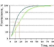 | Maturation curves for TagGFP2 and parental TagGFP.
Dashed lines indicate maturation half-times of 11 min and 18 min for TagGFP2 (dark green curve) and TagGFP (light green curve), respectively. Recording of protein maturation was started when about 7% from their maximal fluorescence has been detected. Time point "0" was defined using an approximation of the beginning of the maturation curves with straight lines.
Data from Subach et al., 2008.
|
|---|
Recommended filter sets and antibodies
The protein can be recognized using Anti-Tag(CGY)FP antibody (Cat.# AB121) or Anti-GFP antibody available from Evrogen.
TagGFP2 can be detected using common fluorescence filter sets for EGFP, FITC, and other green dyes. Recommended Omega Optical filter sets are QMAX-Green, XF100-2, XF100-3, XF115-2, and XF116-2.
Performance and use
TagGFP2 can be easily expressed and detected in a wide range of organisms. Mammalian cells transiently transfected with TagGFP2 expression vectors produce bright fluorescence in 10-12 hrs after transfection. No cytotoxic effects or visible protein aggregation are observed.
TagGFP2 performance in fusions has been demonstrated in the β-actin, α-tubulin and mitochondria-targeting signal models. It can be used in multicolor labeling applications with blue, true-yellow, red, and far-red fluorescent dyes.
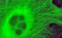
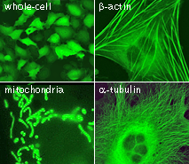 | TagGFP2 expression in transiently transfected mammalian cells.
|
|---|
Available variants and fusions
| Variant | Description | Related vector | Cat.# | Click for image |
|---|
|
|
Humanized TagGFP2
|
TagGFP2 codon usage is optimized for high expression in mammalian cells [Haas et al., 1996], but it can be successfully expressed in many other heterological systems.
|
pTagGFP2-C
|
FP191
|
|
pTagGFP2-N
|
FP192
|
|
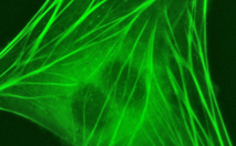
TagGFP2-actin fusion
|
Human β-actin is fused to the TagGFP2 C-terminus. When expressed in mammalian cells, this fusion provides green fluorescent labeling of β-actin in living cells.
|
pTagGFP2-actin
|
FP194
|
|
|
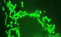
TagGFP2-tubulin fusion
|
Human α-tubulin is fused to the TagGFP2 C-terminus. When expressed in mammalian cells, this fusion provides green fluorescent labeling of α-tubulin in living cells.
|
pTagGFP2-tubulin
|
FP195
|
|
|
TagGFP2-mito fusion
|
A mitochondrial targeting sequence (MTS) is fused to the TagGFP2 N-terminus. MTS was derived from the subunit VIII of human cytochrome C oxidase [Rizzuto et al., 1989; Rizzuto et al., 1995]. When expressed in mammalian cells, this variant provides green fluorescent labeling of mitochondria.
|
pTagGFP2-mito
|
FP197
|
|
References:
-
Haas J, Park EC, Seed B.
Codon usage limitation in the expression of HIV-1 envelope glycoprotein.
Curr Biol. 1996; 6 (3):315-24. / pmid: 8805248
-
Rizzuto R, Brini M, Pizzo P, Murgia M, Pozzan T.
Chimeric green fluorescent protein as a tool for visualizing subcellular organelles in living cells.
Curr Biol. 1995; 5 (6):635-42. / pmid: 7552174
-
Rizzuto R, Nakase H, Darras B, Francke U, Fabrizi GM, Mengel T, Walsh F, Kadenbach B, DiMauro S, Schon EA.
A gene specifying subunit VIII of human cytochrome c oxidase is localized to chromosome 11 and is expressed in both muscle and non-muscle tissues.
J Biol Chem. 1989; 264 (18):10595-600. / pmid: 2543673
-
Subach OM, Gundorov IS, Yoshimura M, Subach FV, Zhang J, Gruenwald D, Souslova EA, Chudakov DM, Verkhusha VV.
Conversion of Red Fluorescent Protein into a Bright Blue Probe.
Chem Biol. 2008; 15 (10):1116-24. doi: 10.1016/j.chembiol.2008.08.006 / pmid: 18940671
-
Xia NS, Luo WX, Zhang J, Xie XY, Yang HJ, Li SW, Chen M, Ng MH.
Bioluminescence of Aequorea macrodactyla, a common jellyfish species in the East China Sea.
Mar Biotechnol (NY). 2002; 4 (2):155-62. / pmid: 14961275
|