|
Far-red fluorescent protein mKate2
- Super bright far-red fluorescence
- Monomeric protein with successful performance in fusions
- Fast maturation, high pH-stability and photostability, low cytotoxicity
- Proven suitability to generate stably transfected cell lines
- Fluorescent signal is easily distinguished from background fluorescence
- Recommended for protein labeling, multicolor applications and whole body imaging
Available variants and fusions
| Variant |
Description |
Related vector |
Cat.# |
Click for image |
|
|
Humanized mKate2
|
mKate2 codon usage is optimized for high expression in mammalian cells [Haas et al., 1996], but it
can be successfully expressed in many other heterological systems.
|
pmKate2-C
|
FP181
|
|
pmKate2-N
|
FP182
|
|
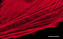
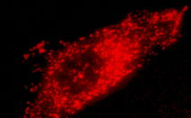
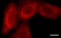
mKate2-actin fusion
|
Human
β-actin
is fused to the mKate2 C-terminus. When expressed in mammalian cells, this fusion provides far-red
fluorescent labeling of
β-actin
in living cells.
|
pmKate2-actin
|
FP184
|
|
|
mKate2-f-mem fusion
|
20 amino acid farnesylation signal from c-Ha-Ras is fused to the mKate2 C-terminus. When expressed in
mammalian cells, this fusion provides far-red fluorescent labeling of plasma membrane.
|
pmKate2-f-mem
|
FP186
|
|
|
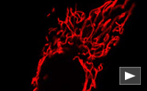
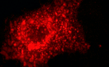
mKate2-mito fusion
|
A mitochondrial targeting sequence (MTS) is fused to the mKate2 N-terminus. MTS was derived from the subunit
VIII of human cytochrome C oxidase [Rizzuto et al., 1989; Rizzuto et al., 1995]. When
expressed in mammalian cells, this variant provides far-red fluorescent labeling of mitochondria.
|
pmKate2-mito
|
FP187
|
|
|
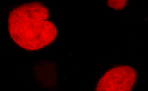
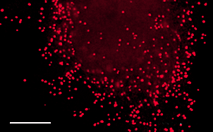
mKate2-H2B fusion
|
Human histone H2B is fused to the mKate2 N-terminus. When expressed in mammalian cells, this fusion provides
far-red fluorescent labeling of histone H2B in living cells.
|
pmKate2-H2B
|
FP311
|
|
|
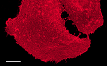
mKate2-lyso fusion
|
Rat Lysosomal Associated Membrane Protein 1 (LAMP-1) is fused to the mKate2 N-terminus. When expressed in
mammalian cells, this fusion provides far-red fluorescent labeling of lysosomes.
|
pmKate2-lyso
|
FP312
|
|
|
mKate2-peroxi fusion
|
Peroxisomal targeting signal [Gould et al., 1989] encoding tripeptide SKL is fused to the 3' end of
mKate2 sequence. This tripeptide targets the fusion protein to the matrix of peroxisomes.
|
pmKate2-peroxi
|
FP313
|
|
|
mKate2-endo fusion
|
Human RhoB GTPase is fused to the mKate2 C-terminus. When expressed in mammalian cells, this fusion provides
far-red fluorescent labeling of vesicles of the endocytic pathway.
|
pmKate2-endo
|
FP314
|
|
|
mKate2-clathrin fusion
|
Human clathrin LCB is fused to the mKate2 C-terminus. When expressed in mammalian cells, this fusion
provides far-red fluorescent labeling of clathrin LCB in living cells.
|
pmKate2-clathrin
|
FP322
|
|
|
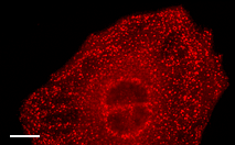
mKate2-ER fusion
|
Signal sequence of calreticulin is fused to the mKate2 N-terminus and the endoplasmic reticulum retention
sequence KDEL is fused to the mKate2 C-terminus. When expressed in mammalian cells, this fusion provides
far-red fluorescent labeling of the lumen of the endoplasmic reticulum.
|
pmKate2-ER
|
FP324
|
|
References:
-
Fliegel L, Burns K, MacLennan DH, Reithmeier RA, Michalak M.
Molecular cloning of the high affinity calcium-binding protein (calreticulin) of skeletal muscle sarcoplasmic reticulum.
J Biol Chem. 1989; 264 (36):21522-8. / pmid: 2600080
-
Gould SJ, Keller GA, Hosken N, Wilkinson J, Subramani S.
A conserved tripeptide sorts proteins to peroxisomes.
J Cell Biol. 1989; 108 (5):1657-64. / pmid: 2654139
-
Haas J, Park EC, Seed B.
Codon usage limitation in the expression of HIV-1 envelope glycoprotein.
Curr Biol. 1996; 6 (3):315-24. / pmid: 8805248
-
Munro S, Pelham HR.
A C-terminal signal prevents secretion of luminal ER proteins.
Cell. 1987; 48 (5):899-907. / pmid: 3545499
-
Rizzuto R, Brini M, Pizzo P, Murgia M, Pozzan T.
Chimeric green fluorescent protein as a tool for visualizing subcellular organelles in living cells.
Curr Biol. 1995; 5 (6):635-42. / pmid: 7552174
-
Rizzuto R, Nakase H, Darras B, Francke U, Fabrizi GM, Mengel T, Walsh F, Kadenbach B, DiMauro S, Schon EA.
A gene specifying subunit VIII of human cytochrome c oxidase is localized to chromosome 11 and is expressed in both muscle and non-muscle tissues.
J Biol Chem. 1989; 264 (18):10595-600. / pmid: 2543673
-
Shcherbo D, Murphy CS, Ermakova GV, Solovieva EA, Chepurnykh TV, Shcheglov AS, Verkhusha VV, Pletnev VZ, Hazelwood KL, Roche PM, Lukyanov S, Zaraisky AG, Davidson MW, Chudakov DM.
Far-red fluorescent tags for protein imaging in living tissues.
Biochem J. 2009; 418 (3):567-74. doi: 10.1042/BJ20081949 / pmid: 19143658
|