|
Near-infrared fluorescent protein NirFP
- Near-infrared fluorescence with emission maximum at 670 nm
- Extremely high photostability
- Fluorescent signal is easily distinguished from background fluorescence
- Recommended for multicolor applications
Performance and use
NirFP can be easily visualized within living tissues. Mammalian cells transiently transfected with NirFP expression vectors produce fluorescence in 48 hrs after transfection. No cytotoxic effects or visible protein aggregation are observed.
Despite its dimeric structure, NirFP can be used in some fusions. However, for protein labeling applications we recommend using specially optimized monomeric TagFPs.
NirFP can be used in multicolor labeling applications with blue, cyan, green, yellow, and red (orange) fluorescent dyes.
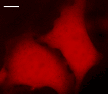 | HeLa cells transiently transfected with pNirFP-N vector.
Widefield Leica AFLX 6000 microscope, 63x objective, after 3 days of incubation. Scale bar, 10 μm. Image from Shcherbo et al., 2010.
|
|---|
References:
-
Shcherbo D, Shemiakina II, Ryabova AV, Luker KE, Schmidt BT, Souslova EA, Gorodnicheva TV, Strukova L, Shidlovskiy KM, Britanova OV, Zaraisky AG, Lukyanov KA, Loschenov VB, Luker GD, Chudakov DM.
Near-infrared fluorescent proteins.
Nat Methods. 2010; 7 (10):827-9. doi: 10.1038/nmeth.1501 / pmid: 20818379
|