
|
||||||||||
|
||||||||||


mKate2
SUPPORTRESOURCES |
|
|||||||||||||||||||

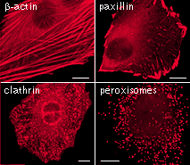 | 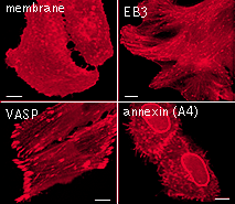 | mKate2 use for protein labeling in mammalian cells.Scale bar represents 10 μm. Images from Shcherbo et al., 2009. |
|---|
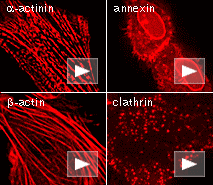 | 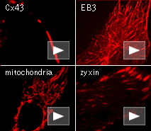 | Live cell imaging of protein dynamics with mKate2.
|
|---|
mKate2 is the superior fluorescent monomeric tag for imaging in living tissues. It has emission maximum at 635 nm optimal for deep imaging of animal tissues, and is more bright, photostable and pH-stable than other cloned far-red monomeric fluorescent proteins.
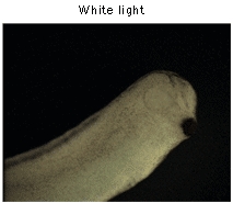
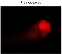
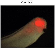
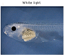
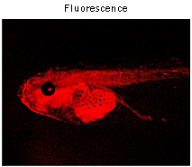
To verify the low toxicity of mKate2 in transgenic animals, mKate2 was expressed under the control of Xanf1 promoter in Xenopus laevis embryos. As expected, bright red fluorescence in the forehead region, including eyes, the forebrain and nasal placodes was observed.
 |  |  | |
|---|---|---|---|
Imaging of mKate2 in Xenopus laevis embryos.Expression of mKate2 under the control of Xanf1 promoter in the transgenic embryos at stage 28 is specifically localized in the forehead region, including eyes, the forebrain and nasal placodes. The embryo is shown from the right side, dorsal to the top and left. Data courtesy of Dr. A. Zaraisky, Institute of Bioorganic Chemistry, RAS (Moscow, Russia). | |||
To compare brightness and maturation rate of TagFP635 and mKate2 in vivo, Xenopus laevis embryos were microinjected with pTagFP635-N and pmKate2-N vectors at the stage of two blastomeres. Living embryos were photographed from the animal pole side at the middle gastrula stages (10.5 hours after fertilization). As expected, embryos microinjected with pmKate2-N demonstrated superior brightness.
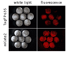 | Comparison of TagFP635 and mKate2 in developing Xenopus laevis embryos.Data courtesy of Dr. A. Zaraisky, Institute of Bioorganic Chemistry, RAS (Moscow, Russia). |
|---|
In addition, to test in an embryonic model the performance of mKate2 in a targeting protein fusion, we generated transgenic Xenopus laevis embryos bearing a CMV-mKate2-zyxin fusion construct. Despite quite extensive and ubiquitous expression of mKate2-zyxin under the control of the CMV promoter, these embryos appear normal and healthy indicating that mKate2 exerts a low toxic effect on living cells in transgenic organisms.
 |  | Expression of mKate2-zyxin in Xenopus laevis embryo.Data courtesy of Dr. A. Zaraisky, Institute of Bioorganic Chemistry, RAS (Moscow, Russia). |
|---|
mKate2 can be used in multicolor labeling applications with blue, cyan, green, yellow, and red (orange) fluorescent dyes. High pH-stability with pKa=5.4 makes it possible to use mKate2 for imaging in acidic organelles, such as late and recycling endosomes and lysosomes.
References:
- Shcherbo D, Murphy CS, Ermakova GV, Solovieva EA, Chepurnykh TV, Shcheglov AS, Verkhusha VV, Pletnev VZ, Hazelwood KL, Roche PM, Lukyanov S, Zaraisky AG, Davidson MW, Chudakov DM. Far-red fluorescent tags for protein imaging in living tissues. Biochem J. 2009; 418 (3):567-74. doi: 10.1042/BJ20081949 / pmid: 19143658
|
Copyright 2002-2023 Evrogen. All rights reserved. Evrogen JSC, 16/10 Miklukho-Maklaya str., Moscow, Russia, Tel +7(495)988-4084, Fax +7(495)988-4085, e-mail:evrogen@evrogen.com |