|
Photoactivatable red fluorescent protein PA-TagRFP
- Monomer, successful performance in fusions
- Non-fluorescent before photoactivation
- Irreversible photoactivation to a red fluorescent form by UV-violet light irradiation
- High brightness and photostability
- Recommended for super-resolution imaging
Performance and use
PA-TagRFP can be easily expressed and detected in a wide range of organisms. Mammalian cells transiently transfected with PA-TagRFP expression vectors produce bright fluorescence upon UV-activation of PA-TagRFP in 10-12 hrs after transfection. No cytotoxic effects or visible protein aggregation are observed.
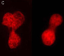
 | PA-TagRFP use for cell labeling.
Live HeLa cells transiently transfected with the PA-TagRFP-C expression vector were imaged during the photoactivation.
|
|---|
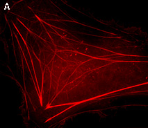
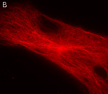
PA-TagRFP performance in protein fusions has been demonstrated in β-actin, α-tubulin, histone H2B and other models.
PA-TagRFP use in PALM imaging techniques
High brightness, photostability and absence of initial fluorescence signal from PA-TagRFP make it a protein tag of choice for super resolution two-color PALM/single-particle tracking PALM imaging techniques. The excellent performance of PA-TagRFP in two-color single-particle tracking PALM experiments was demonstrated for several PA-TagRFP-tagged and PAGFP-tagged fusions in live COS-7 cells [Subach et al., 2010].
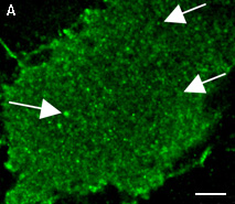
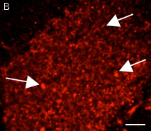
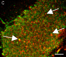
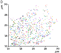
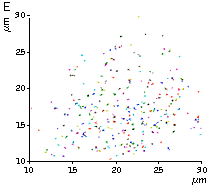
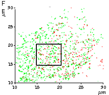
An example for the tracking of PA-TagRFP-tagged epidermal growth factor receptor (EGFR-PATagRFP) and PAGFP-tagged vesicular stomatitus virus G protein tsO45 (VSVG-PAGFP) in live COS-7 cells by two-color single-particle tracking PALM is shown below.
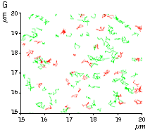 |
(G) A zoomed view of the region indicated by the square in (F).
|
|---|
References:
-
Subach FV, Patterson GH, Renz M, Lippincott-Schwartz J, Verkhusha VV.
Bright monomeric photoactivatable red fluorescent protein for two-color super-resolution sptPALM of live cells.
J Am Chem Soc. 2010; 132 (18):6481-91. doi: 10.1021/ja100906g / pmid: 20394363
|