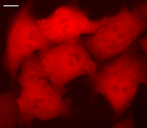
HeLa cells transiently transfected with pTurboFP650-N vector.
Widefield Leica AFLX 6000 microscope, 63x objective, after 3 days of incubation. Scale bar, 10 μm. Image from Shcherbo et al., 2010.
TurboFP650 can be easily visualized within living tissues. Mammalian cells transiently transfected with TurboFP650 expression vectors produce bright fluorescence in 14 hrs after transfection. No cytotoxic effects or visible protein aggregation are observed.
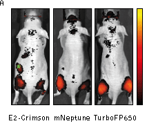
Superior performance of TurboFP650 in whole-body imaging was demonstrated using mouse xenograft model. HEK 293T cells transiently transfected with a plasmids encoding either TurboFP635, TurboFP650, NirFP, mNeptune or E2-Crimson were injected intramuscularly into the gluteal region of mice. The cells were co-transfected with firefly luciferase plasmid to normalize the transfection efficiency and total numbers of injected cells. Imaging of cell implants and quantification at various emission wavelengths showed higher fluorescence from TurboFP650 at two excitation wavelengths.
Despite its dimeric structure, TurboFP650 can be used in some fusions. However, for protein labeling applications we recommend using specially optimized monomeric TagFPs.
TurboFP650 can be used in multicolor labeling applications with blue, cyan, green, yellow, and red (orange) fluorescent dyes.
| HeLa cells transiently transfected with pTurboFP650-N vector.Widefield Leica AFLX 6000 microscope, 63x objective, after 3 days of incubation. Scale bar, 10 μm. Image from Shcherbo et al., 2010. |
|---|
 |  |  | |
|---|---|---|---|
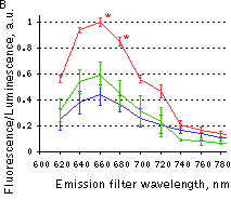
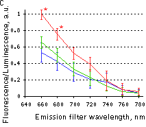
Whole-mouse imaging with IVIS Spectrum system (Caliper).(A) Representative fluorescence reflectance images (excitation filter, 605/30 nm and emission filter, 660/20 nm) of mice injected intramuscularly with HEK 293T cells expressing E2-Crimson, mNeptune or TurboFP650. Green asterisk denotes background fluorescence in mice injected with E2-Crimson cells. The color bar indicates radiant efficiency ×10-6; minimum is 0.001, and maximum is 0.006. (B,C) Fluorescence efficiency from cell implants imaged with 570/30 nm (B) or 605/30 nm (C) excitation filters and various emission filters, normalized to photons from firefly luciferase to control for transfection efficiency and numbers of implanted cells. Means ± s.e.m. are shown (n = 6-10 per point). *P < 0.05 (Student's t-test) for TurboFP650 relative to other proteins. Red line – TurboFP650, green line – mNeptune, blue line – E2-Crimson. Images and data from Shcherbo et al., 2010. | |||