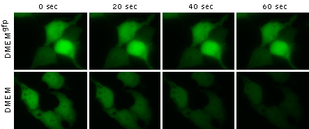
Long-term fluorescent microscopy of EGFP in live HEK293T cells maintained in DMEM or DMEMgfp.
30 min before starting imaging experiment replace the cell culture medium with DMEMgfp-2 as is or supplemented with rutin at a final concentration of 20 mg/l. Always prepare a fresh solution of rutin in DMEMgfp-2! The replacement should be performed under sterile conditions.
For long experiments DMEMgfp-2 can be supplemented with L-glutamine, penicillin, streptomycin and fetal bovine serum.
| Long-term fluorescent microscopy of EGFP in live HEK293T cells maintained in DMEM or DMEMgfp.
|
|---|
 |  |  | |
|---|---|---|---|
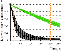
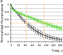
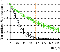
Influence of cell medium on photostability of fluorescent proteins.(A) – EGFP, (B) AcGFP1, (C) TagGFP2. Graph shows normalized bleaching curves of fluorescent proteins in live HEK293 cells maintained in DMEM (black lines), or DMEMgfp (green lines). Standard deviations (n = 15-20 cells) are shown.
| |||
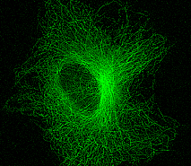 | 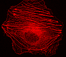 | HeLa cells transfected with fluorescent protein-tagged α-tubulin or β-actin had a normal cytoskeleton after 5-day culture in DMEMgfp.
Fluorescence microscopy of TagGFP2- |
|---|
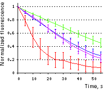 | Influence of rutin on EGFP photobehavior.Bleaching of green fluorescence in EGFP-expressing live HEK293 cells maintained in DMEM (red), DMEM with rutin (blue), DMEMgfp-2 (violet), or DMEMgfp-2 with rutin (green). Green fluorescence intensities in individual cells are background subtracted and normalized to maximum (initial) value in each cell. Standard deviation values (n = 15–20 cells in a representative experiment out of five independent experiments) are shown. Data from Bogdanov et al., 2012. |
|---|