
|
||||||||||
|
||||||||||


TurboFP650
SUPPORTRESOURCES |
|
||||||||||

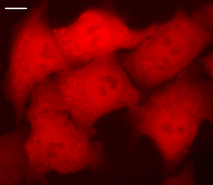 | HeLa cells transiently transfected with pTurboFP650-N vector.Widefield Leica AFLX 6000 microscope, 63x objective, after 3 days of incubation. Scale bar, 10 μm. Image from Shcherbo et al., 2010. |
|---|
 |  |  | |
|---|---|---|---|
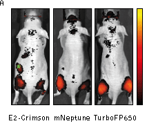
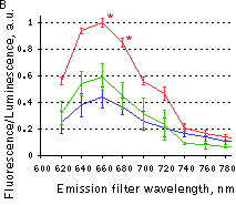
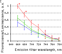
Whole-mouse imaging with IVIS Spectrum system (Caliper).(A) Representative fluorescence reflectance images (excitation filter, 605/30 nm and emission filter, 660/20 nm) of mice injected intramuscularly with HEK 293T cells expressing E2-Crimson, mNeptune or TurboFP650. Green asterisk denotes background fluorescence in mice injected with E2-Crimson cells. The color bar indicates radiant efficiency ×10-6; minimum is 0.001, and maximum is 0.006. (B,C) Fluorescence efficiency from cell implants imaged with 570/30 nm (B) or 605/30 nm (C) excitation filters and various emission filters, normalized to photons from firefly luciferase to control for transfection efficiency and numbers of implanted cells. Means ± s.e.m. are shown (n = 6-10 per point). *P < 0.05 (Student's t-test) for TurboFP650 relative to other proteins. Red line – TurboFP650, green line – mNeptune, blue line – E2-Crimson. Images and data from Shcherbo et al., 2010. | |||
References:
- Shcherbo D, Shemiakina II, Ryabova AV, Luker KE, Schmidt BT, Souslova EA, Gorodnicheva TV, Strukova L, Shidlovskiy KM, Britanova OV, Zaraisky AG, Lukyanov KA, Loschenov VB, Luker GD, Chudakov DM. Near-infrared fluorescent proteins. Nat Methods. 2010; 7 (10):827-9. doi: 10.1038/nmeth.1501 / pmid: 20818379
|
Copyright 2002-2023 Evrogen. All rights reserved. Evrogen JSC, 16/10 Miklukho-Maklaya str., Moscow, Russia, Tel +7(495)988-4084, Fax +7(495)988-4085, e-mail:evrogen@evrogen.com |