Kindling red fluorescent protein KFP-Red
- Reversible or irreversible photoactivation
- Activated by green light that does not damage cells and tissues
- Quenching by blue light
- Recommended for tracking cells and cellular organelle movements
Performance and use
KFP-Red was successfully expressed and tested in various experimental models, including bacteria, Xenopus embryo, and cultured mammalian cells. Like other Anthozoa GFP-like proteins, KFP-Red is a tetramer. This restricts the wide use of KFP-Red as a fusion partner for cellular proteins.
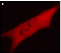 | 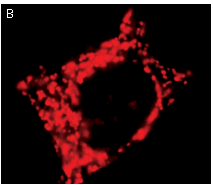 | KFP-Red use for cell (A) and mitochondria labeling (B).
Fluorescent microscopic images were obtained after reversible kindling of KFP-Red in HeLa cells.
|
|---|
Reversible or irreversible kindling: Depending on the kindling light intensity KFP-Red can be photoactivated reversibly or irreversibly allowing the monitoring of both short- and long-term cell processes.
A reversibly kindled KFP-Red relaxes to the initial non-fluorescent form (half-life 50 seconds), or can be quenched instantly by blue light (430-490 nm). Reversible kindling results in about 70 times increase of the red fluorescence intensity comparing to unkindled protein. Reversible kindling and quenching can be repeated many times.
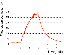 |  | KFP-Red reversible kindling and relaxation.
(A) Kindling and relaxation kinetics. Zero time is set at the commencement of irradiation with kindling light (532 nm laser light, 1% power). Kindling irradiation was stopped after 2 min. (B) Reversible photoactivation of KFP-Red in E. coli. The round-shaped part of the E. coli colony exp-ressing KFP-Red was irreversibly kindled with intense green light. This region fluoresces brightly, while its absorption is low. After several minutes, the kindled protein relaxed to the non-fluorescent state, while its absorption recovered.
|
|---|
An irreversibly kindled KFP-Red gives stable red fluorescence which is at least 35 times brighter than that of the protein before kindling. An irreversibly kindled KFP-Red remains stable and brightly fluorescent for more than 72 hrs in living cells and for at least a year in protein samples.
An irreversibly kindled KFP-Red can be partially quenched by blue light, but then it restores its brightness within several minutes. Therefore, in some applications, blue light can be used to quench a reversibly kindled KFP-Red, whereas an irreversibly kindled KFP-Red remains fluorescent.
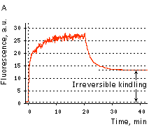 |  | KFP-Red irreversible kindling.
(A) Kindling kinetics. Zero time is set at the commencement of irradiation with kindling light (532 nm laser light, 20% power). Kindling irradiation was stopped after 20 min. (B) Irreversibly kindled (red line) and "unkindled" (blue line) KFP-Red fluorescence spectra and brightness ratio. The photo shows intact and irreversibly kindled KFP-Red samples after a year of incubation at room temperature.
|
|---|
Application of KFP-Red to track cell migration was demonstrated using embryonic fate mapping as an example. Xenopus embryos were taken at the stage of two blastomeres and KFP-Red mRNA was microinjected into the animal poles of both blastomeres. At the early neurula stage, a round-shaped group of cells within the neural plate was kindled irreversibly. Irradiated cells became brightly fluorescent and their migration in the developing embryo was monitored. Longitudinal extension accompanied by transversal convergence of the labeled group of cells was visible after the first two hours after kindling. At the end of neurulation, the labeled spot appeared as a thin stripe on the surface of the left neural fold.
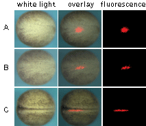 | Monitoring of cell migration during Xenopus neural plate development using KFP-Red.
(A) At the early neurula stage, a round-shaped group of cells within the neural plate was irreversibly "kindled"; (B) longitudinal extension of the labeled group of cells after two hours after kindling; (C) thin stripe of the labeled cells at the end of neurulation.
Experimental data were presented by Dr. A. Zaraisky, Institute of Bioorganic Chemistry, RAS (Moscow, Russia).
|
|---|
KFP-Red suitability for tracking movement of cell organelles was demonstrated on PC12 cells transfected with a mitochondria-targeted KFP-Red expressing vector. After 25 hours of incubation, mitochondria remained non-fluorescent (no kindling observed) upon irradiation using a 1% power scanning green laser (HeNe laser line 543 nm, 1 mW, once per 10 seconds; the number of scans is not limited). After several scans with a 5-10% power laser, mitochondria became brightly fluorescent and were observed using a 1% power laser for several minutes. Brief irradiation (about 20 seconds in fast mode) with a 30% power green laser light induced irreversible kindling of KFP-Red in mitochondria within the irradiated field. Irreversibly kindled mitochondria were monitored.
 | Monitoring of mitochondria movement in PC12 cells using KFP-Red (movie).
|
|---|
KFP-Red diffusion within an eukaryotic cell was also demonstrated. HeLa cells were transfected with pKindling-Red-N vector. After 24 hours of incubation they were observed in the Zeiss LSM 510 confocal microscope. Almost no fluorescence could be observed upon irradiation by 1% power of 1mW 543 nm laser. The upper part of the cell was shortly (about 5-10 seconds) irradiated by 100% power 543 nm laser, causing kindling of the KFP-Red. Starting immediately after kindling, diffusion of the activated protein was observed using 1% power laser as an excitation light source.
 | KFP-Red diffusion within eukaryotic cell (movie).
|
|---|
See additional examples of KFP-Red use at www.olympusconfocal.com
|